Histopathological diagnosis of small cell lung cancer through the biopsy of spleen metastases under ultrasound examination
Marek Chorazy
Department of Clinical Oncology and Internal Medicine, St.Leszczynski Hospital Raciborska 27. 40-074 Katowice, Poland
Abstract:
Mostly, the pathological diagnosis is conducted on the basis of examination of a tumor. Also, the similar results are obtained during the process of collecting specimens either surgically or during a biopsy. Occasionally, the diagnosis involves collecting a sample during a biopsy of metastasis to the other organs . Splenic metastases are very rare and are mostly diagnosed at the terminal phase of the disease or at the time of autopsy. The histological examination if is done, is made prevalently by splenectomy. A few described reports of splenic percutaneous biopsies in the diagnosis of splenic metastasis are fragmentary and very poor. It is connected with the fact , that splenic metastasis are secondary lesions, generally being found in patients with other metastases (particularly situated in the liver).
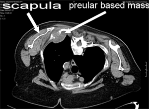
A woman patient (aged 70) was sent from a hospital in Częstochowa for a further diagnosis of a left lung tumor lesion discovered at an X-ray examination. A computer tomography (CT) scan has shown a chest infiltration of a pulmonary tissue (fig 1), most probably neoplasmatic, situated under the left scapula. A CT guided percutaneus biopsy was performed twice without positive results because the needle didn’t reach the tumor. in the subscapular region.
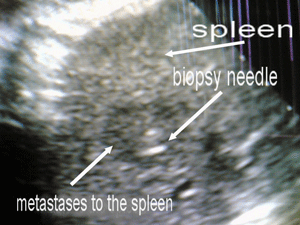
In this case we decided to look for secondary lesions in other organs. A ultrasound examination of the abdomen revealed a pathologic mass in the spleen. We made a ultrasound guided (free hand technique) fine - needle biopsy of the lesion (fig 2). The biopsy was positive and a diagnosis of small-cell cancer, which probably originated in the lungs was established. The patient was treated with chemotherapy (cisplatinum + etoposidum) in the oncology department of our hospital.
In the case of this patient, pathologic diagnosis was made during a biopsy of isolated spleen metastasis, which is conducted occasionally. The case presented above is an example that metastasis can occur in every organ of human body. Sometimes it can be isolated in the least probably organs we could expect. And when such organ is available for the biopsy we are able to establish pathological diagnosis.
Figure legends:
Fig 1. CT - lung tumor under the left scapula
Fig 2. Ultrasound – puncture needle in metastases to spleen